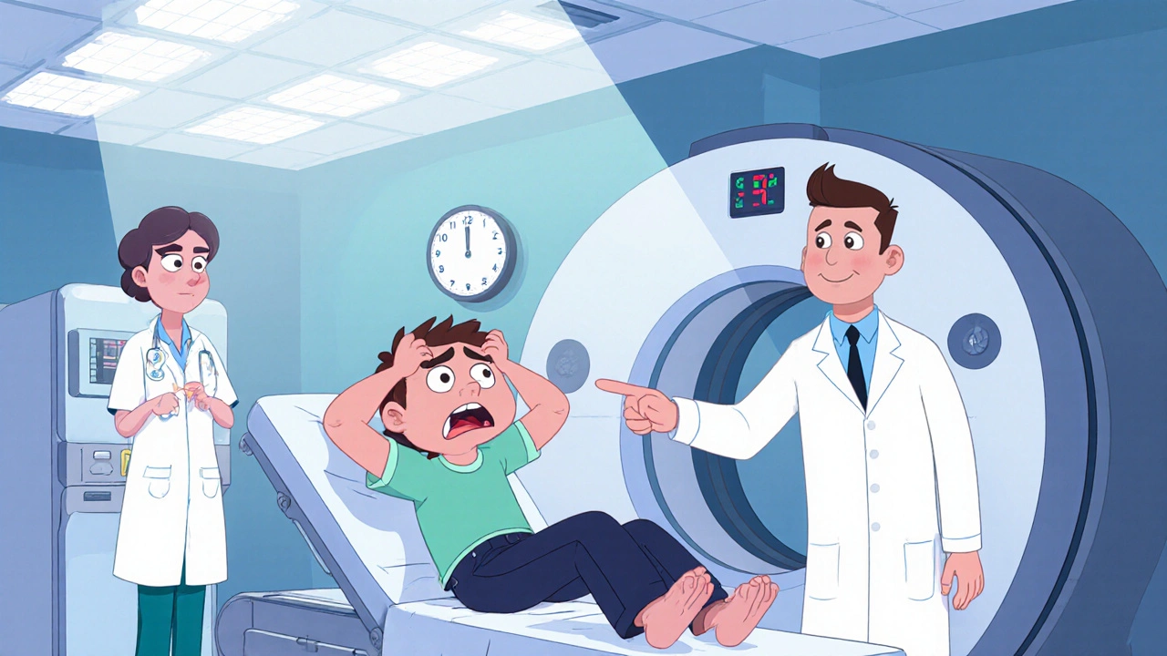
MRI (Magnetic Resonance Imaging)
When working with MRI, a non‑invasive scan that uses magnetic fields and radio waves to create detailed pictures of the inside of the body. Also known as magnetic resonance imaging, it lets doctors see soft tissue, brain, joints, and many other structures without radiation. Contrast agent a special dye injected to highlight blood vessels and lesions often improves the clarity of the images, especially in functional MRI a technique that maps brain activity by tracking blood‑flow changes. In the world of radiology the medical specialty that interprets imaging studies, MRI has become a go‑to tool for diagnosing everything from torn ligaments to brain tumors. If you’re wondering why MRI is so popular, the answer lies in its versatility and safety.
One of the core strengths of MRI is its ability to change the pulse sequence, which essentially swaps the way the scanner talks to the body. For instance, T1‑weighted imaging highlights fat and provides clear anatomical detail shines when doctors need a crisp view of brain structure or spinal cord anatomy. On the flip side, diffusion weighted imaging captures the movement of water molecules to spot acute stroke or tumor infiltration helps emergency teams act fast. The scanner also demands a strong magnet—usually 1.5 or 3 Tesla—to generate the signal, which means safety checks are a must, especially for patients with pacemakers or metal implants. Knowing the right protocol, choosing whether to add a contrast agent, and understanding which sequence fits the clinical question are all part of the decision‑making chain that makes MRI such a powerful diagnostic ally.
What you’ll find below
Below is a curated list of articles that connect MRI to real‑world medical scenarios. You’ll see how imaging guides treatment choices for conditions covered in our drug guides—from monitoring chemotherapy response to checking for joint inflammation after taking anti‑inflammatories. Whether you’re a patient curious about what an MRI report means, a caregiver trying to understand why a contrast injection is recommended, or a health‑professional looking for concise overviews, the posts ahead give practical tips, safety pointers, and clear explanations. Dive in to see how MRI fits into the broader picture of modern healthcare.
Why Neuroimaging is Critical for Diagnosing Subarachnoid Hemorrhage
Learn why rapid neuroimaging-CT, MRI, and angiography-is essential for diagnosing subarachnoid hemorrhage, guiding treatment, and improving outcomes.
More