SAH Imaging Decision Tool
Determine Optimal Imaging Pathway for SAH Diagnosis
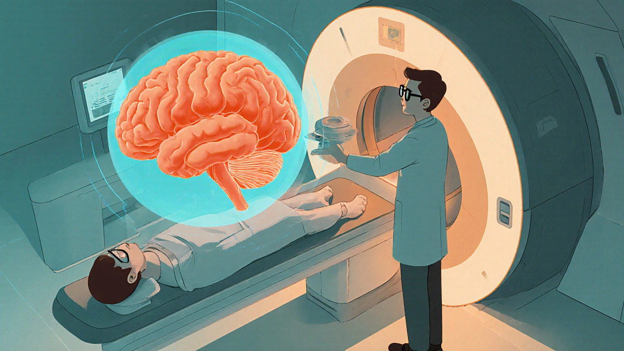
When a patient arrives with a sudden, severe headache, doctors need rapid, reliable tools to confirm or rule out a Subarachnoid Hemorrhage is bleeding into the space between the brain and its covering membranes, often caused by a ruptured aneurysm. The cornerstone of that decision‑making process is neuroimaging, a suite of techniques that let clinicians see inside the skull without surgery.
Why Early Diagnosis Saves Lives
Subarachnoid hemorrhage (SAH) carries a mortality rate of 25‑30 % within the first month, and half of survivors are left with lasting neurological deficits. Prompt detection enables:
- Immediate blood‑pressure control to prevent re‑bleeding.
- Early aneurysm repair-either endovascular coiling or surgical clipping.
- Targeted monitoring for complications like cerebral vasospasm.
- Informed prognostication using established clinical scales.
Every minute counts, which is why emergency departments rely on fast, sensitive imaging.
CT Scan: The First‑Line Workhorse
A non‑contrast CT Scan is computed tomography, an X‑ray based technique that produces cross‑sectional images of the brain in seconds. Its strengths for acute SAH include:
- Detection of blood as hyperdense (bright) areas in the subarachnoid space within the first 6 hours.
- Wide availability in most hospitals, including rural emergency units.
- Rapid acquisition-often under one minute of scanning time.
Modern 64‑slice scanners can spot even 1 mm of blood, pushing sensitivity above 95 % in the first 12 hours. However, CT’s ability declines after 24 hours as clot density normalizes.
MRI: The Detail‑Oriented Detective
When CT results are equivocal or the bleed is delayed, a MRI is magnetic resonance imaging, which uses magnetic fields and radio waves to generate high‑resolution images of soft tissue with sequences like FLAIR and T2* that highlight older blood products.
- FLAIR (Fluid‑Attenuated Inversion Recovery) suppresses CSF signals, making subtle subarachnoid blood appear bright.
- SWI (Susceptibility‑Weighted Imaging) is especially good at detecting hemosiderin from prior bleeds.
- MRI also evaluates the surrounding brain for ischemia, infarcts, or edema-information vital for surgical planning.
The trade‑off is longer scan time (usually 20-30 minutes) and limited access in smaller centers.
Digital Subtraction Angiography (DSA): Mapping the Source
Even the best non‑invasive images can miss a tiny aneurysm. Digital Subtraction Angiography is an invasive X‑ray procedure that injects contrast into cerebral vessels, producing dynamic, high‑resolution vascular maps. DSA remains the gold standard for:
- Identifying the exact location, size, and neck morphology of an aneurysm.
- Guiding endovascular treatment during the same session.
- Detecting vasospasm or arterial dissection not visible on CT or MRI.
Because it carries a small risk of stroke (≈0.5 %) and requires a specialized cath lab, DSA is typically reserved for cases where non‑invasive imaging is inconclusive or when treatment is imminent.

Scoring Systems and Imaging Interpretation
Clinicians blend imaging findings with clinical scales to predict outcomes:
- Hunt and Hess Scale grades patients from I (asymptomatic) to V (deep coma), reflecting neurological status.
- Fisher Scale categorizes the amount of blood seen on CT, ranging from 1 (minimal) to 4 (diffuse or intraventricular).
- Glasgow Coma Scale quantifies consciousness, influencing urgent imaging decisions.
For example, a Fisher Grade 3 bleed (thick cisternal clot) combined with a Hunt‑Hess Grade III patient signals a high risk of vasospasm, prompting early transcranial Doppler monitoring.
Practical Emergency Department Workflow
A typical SAH work‑up looks like this:
- Patient presents with “worst headache of life.”
- Rapid neurological exam → if GCS < 15 or focal deficits, proceed immediately to CT Scan.
- If CT is negative but suspicion remains (e.g., neck stiffness, delayed presentation), order MRI with FLAIR/SWI.
- Positive imaging → consult Radiologist is medical doctor specialized in interpreting imaging studies for precise aneurysm characterization.
- Based on size and location, the neurosurgical team decides between endovascular coiling (often done in the same DSA suite) or surgical clipping.
- Post‑procedure, initiate nimodipine therapy and monitor for Cerebral Vasospasm is constriction of cerebral arteries that can cause delayed ischemia using daily transcranial Doppler.
This algorithm balances speed with diagnostic accuracy, ensuring patients get the right treatment at the right time.
Common Pitfalls and How to Avoid Them
- Missing Small Aneurysms: Use thin‑slice CT angiography (CTA) or DSA when initial non‑contrast CT is negative but clinical suspicion stays high.
- Misreading Calcifications as Blood: Dual‑energy CT can differentiate calcium from hemorrhage.
- Delay in Imaging: Aim for CT within 30 minutes of arrival; each hour of delay can increase re‑bleeding risk by ~4 %.
- Over‑reliance on One Modality: Combine CT (for acute bleed) with CTA or MRI (for aneurysm morphology) for a comprehensive view.

Future Directions: Advanced Imaging and AI
Artificial intelligence algorithms are being trained on thousands of SAH cases to flag subtle blood on CT and suggest optimal vascular imaging pathways. Additionally, 7‑Tesla MRI promises even finer detection of micro‑hemorrhages and vessel wall inflammation, potentially identifying patients at risk before rupture.
While these technologies are still emerging, they hint at a future where neuroimaging not only diagnoses SAH faster but also predicts it.
Key Takeaways
- Early neuroimaging reduces mortality and improves functional outcomes in subarachnoid hemorrhage.
- Non‑contrast CT is the fastest first‑line test; MRI adds sensitivity for delayed or subtle bleeds.
- Digital Subtraction Angiography remains essential for aneurysm mapping and treatment planning.
- Integrating imaging findings with clinical scales (Hunt‑Hess, Fisher, GCS) guides prognosis and management.
- Emerging AI‑driven tools may soon streamline detection and decision‑making.
Frequently Asked Questions
Can a subarachnoid hemorrhage be missed on a CT scan?
Yes, especially if the scan is performed more than 24 hours after symptom onset. Blood density fades, making subtle hemorrhage hard to spot. In such cases, an MRI with FLAIR or a CTA is recommended.
Is MRI safe for patients with a possible aneurysm?
MRI does not use ionizing radiation, so it’s safe for most patients. However, if a metallic implant is present, the scan may be contraindicated. The choice between MRI and CTA depends on availability and clinical urgency.
When is Digital Subtraction Angiography mandatory?
DSA is mandatory when non‑invasive imaging fails to clearly identify the aneurysm’s size, neck, or exact location, or when immediate endovascular treatment is being considered.
How quickly should treatment start after a SAH diagnosis?
Ideally within 24 hours of rupture. Early securing of the aneurysm (clipping or coiling) dramatically lowers re‑bleeding risk and improves survival.
What role does the Fisher Scale play in imaging?
The Fisher Scale grades the amount of blood seen on CT, helping predict the likelihood of vasospasm. Higher grades (3‑4) signal a need for aggressive monitoring and prophylactic therapy.
Comparison of Common Imaging Modalities for SAH
| Feature | CT Scan (Non‑contrast) | MRI (FLAIR/SWI) | Digital Subtraction Angiography |
|---|---|---|---|
| Acute bleed detection | Excellent (90‑95 % within 6 h) | Good, but slower | Gold standard for vascular detail |
| Time to result | 1‑2 min | 20‑30 min | 30‑45 min (including prep) |
| Aneurysm size/neck visualization | Limited (requires CTA) | Moderate (high‑res 3D MRI possible) | High‑resolution, 0.1 mm |
| Invasiveness | Non‑invasive | Non‑invasive | Invasive (catheter, contrast) |
| Complication risk | Radiation exposure | None (magnetic fields) | Stroke ~0.5 %, contrast nephropathy |
| Best use case | Initial assessment within hours of symptom onset | Late presentation or equivocal CT | Pre‑treatment planning or when CTA is inconclusive |
Choosing the right modality hinges on timing, availability, and the clinical question at hand. In practice, most hospitals start with a rapid CT, then follow up with MRI or DSA as needed.


Benedict Posadas
Yo! CTs are the real MVP in SAH 🚀
Jai Reed
Non‑contrast CT remains the fastest and most reliable first‑line tool for detecting acute subarachnoid hemorrhage. Its sensitivity exceeds 95 % within the first 12 hours, making it indispensable in emergency settings. However, clinicians must recognize the decreasing sensitivity after 24 hours and follow up with MRI or CTA when suspicion persists. Early imaging directly influences treatment decisions and improves patient outcomes.
WILLIS jotrin
It’s fascinating how a few seconds of scanning can mean the difference between life and death. While technology gives us speed, the human judgment in interpreting those images is equally crucial. We often balance the urgency with the need for detailed vascular mapping, especially when the bleed is subtle. The interplay of CT and subsequent modalities showcases a layered diagnostic approach. In the end, it’s a dance between machines and minds.
Joanne Ponnappa
CT scans are quick, but remember they expose patients to radiation. 🧭 It’s wise to limit repeat scans and consider MRI when feasible.
Emily Collins
The shadows on those slices whisper the tragedy of a ruptured vessel, a silent scream captured in grayscale, urging us to act before the next wave of blood drowns the brain.
Harini Prakash
From a broader perspective, integrating AI‑driven detection algorithms with traditional CT could further reduce missed cases. In many low‑resource settings, rapid CT still saves lives, but we should push for equitable access to MRI and DSA. Collaboration between radiologists, neurologists, and engineers is key to advancing these tools.
Rachael Turner
Imaging is the lighthouse that guides us through the fog of an uncertain diagnosis. When a patient screams about the worst headache of their life the clock starts ticking and every minute without a clear picture feels like a lost opportunity. The CT scan bursts onto the scene like a sprinter, delivering a view of the brain in under a minute and showing bright blood that could not be ignored. Its speed is its greatest asset but also its Achilles heel because after the first day the hyperdensity fades and the bleed can hide. That is where MRI steps in like a detective with a magnifying glass, using FLAIR and SWI sequences to pull out the subtle clues that CT missed. The longer scan time is a price some hospitals are willing to pay for that extra detail especially when the patient’s presentation is delayed. Digital Subtraction Angiography, on the other hand, is the artisan’s tool carving out the exact shape and neck of the aneurysm. It is invasive and not without risk but the reward is a roadmap that can be used instantly for coiling or clipping. The clinical scales such as Hunt‑Hess and Fisher are not just numbers they are lenses that focus the imaging findings into a prognosis. A high Fisher grade on the CT tells us to watch for vasospasm like a storm brewing over the horizon. The integration of these images with the patient’s neurological exam creates a synergy that drives treatment decisions. In many community hospitals the first CT is the only modality available and it often saves lives simply by ruling out other emergencies. Yet we must keep pushing for wider access to MRI and DSA so that no subtle bleed goes unnoticed. The future promises AI algorithms that will flag even the faintest bleed on a CT slice before the radiologist even looks at it. Imagine a world where a computer whispers “possible SAH” at the moment the scan is completed and the team moves to secure the aneurysm within hours. Until then the old mantra still holds true – fast, accurate imaging equals better outcomes for patients facing subarachnoid hemorrhage.
Vin Alls
The cascade you described is spot‑on – it’s like watching a symphony where CT drums the beat, MRI strings the nuance, and DSA solos the finale. Adding AI into that orchestra could turn a good performance into a masterpiece, catching those stealthy bleeds before they wreak havoc. Let’s keep feeding the algorithms with diverse data so they learn the full spectrum of SAH presentations.
Tiffany Davis
I agree that interdisciplinary collaboration is essential for improving SAH care. Sharing protocols and data across institutions can help standardize imaging pathways and reduce delays.