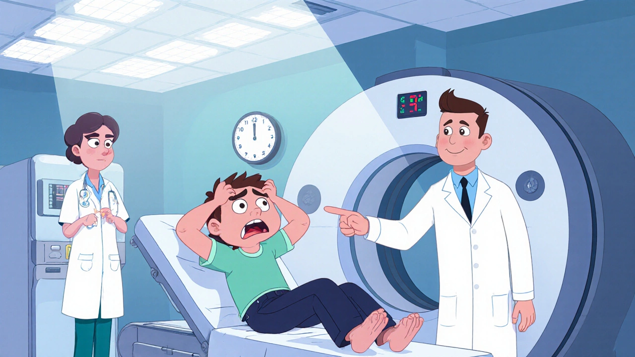
Neuroimaging: Understanding Brain Imaging Techniques
When working with neuroimaging, the use of medical imaging to visualize the structure and function of the nervous system. Also known as brain imaging, it helps doctors diagnose, monitor, and research neurological conditions. MRI, magnetic resonance imaging, provides detailed soft‑tissue contrast without radiation and CT, computed tomography, uses X‑rays to capture fast cross‑sectional images are the workhorses of structural neuroimaging. Together they form a toolkit that covers most anatomical questions. In addition, functional methods such as PET, positron emission tomography, maps metabolic activity with radioactive tracers add a layer of physiological insight. These modalities let clinicians see both the hardware and the software of the brain.
Neuroimaging encompasses MRI and CT scans, which means you can choose the right tool based on speed, detail, and safety. MRI requires strong magnetic fields, so patients with implants need special screening, while CT requires ionizing radiation, making it ideal for acute emergencies like head trauma. The high spatial resolution of MRI helps spot tiny lesions in multiple sclerosis, whereas CT shines when you need a quick view of bleeding after a stroke. Both techniques share a common goal: they turn invisible brain structures into clear pictures doctors can act on.
Functional neuroimaging goes a step further. PET requires radioactive tracers that highlight glucose metabolism, which is useful for detecting early Alzheimer changes. Another key player is functional MRI, often called fMRI, functional magnetic resonance imaging, which tracks blood‑oxygen‑level changes during brain activity. fMRI informs researchers which regions light up when you solve a puzzle, while PET can reveal how a tumor steals sugar from surrounding tissue. Together, these modalities provide a two‑dimensional view of structure and a three‑dimensional view of function, a combo that guides treatment plans for epilepsy, depression, and brain tumors.
In clinical practice, neuroimaging answers real‑world questions. When a patient arrives with sudden weakness, a CT scan quickly shows whether a bleed or clot is present, allowing emergency teams to act within the golden hour. For chronic conditions like Parkinson’s, MRI helps assess iron deposition, and PET can track dopamine loss over time. Even psychiatric disorders benefit: fMRI studies reveal altered connectivity in anxiety, guiding personalized therapy. The link between imaging findings and patient outcomes makes neuroimaging a cornerstone of modern neurology and neurosurgery.
Research is pushing the envelope with newer techniques. Diffusion tensor imaging (DTI) maps white‑matter pathways, revealing hidden damage after concussions. Artificial‑intelligence algorithms now scan thousands of brain images in seconds, spotting patterns that escape the human eye. Hybrid scanners combine PET and MRI into a single machine, delivering structural and metabolic data side by side. These advances are expanding what neuroimaging can do—from early disease detection to monitoring how drugs reshape brain activity.
Below you’ll find a curated list of articles that dive deeper into each of these topics. Whether you’re looking for side‑by‑side drug comparisons, safety guidelines, or practical tips for balancing medication with daily life, the posts map directly onto the imaging concepts we’ve just covered. Explore the collection to see how neuroimaging informs treatment choices, research directions, and everyday health decisions.
Why Neuroimaging is Critical for Diagnosing Subarachnoid Hemorrhage
Learn why rapid neuroimaging-CT, MRI, and angiography-is essential for diagnosing subarachnoid hemorrhage, guiding treatment, and improving outcomes.
More
